in vivo MN Test

In toxicology, cytostatic effects of the test compound are measured by assessing the ratio of mono-, bi- and multi-nucleated cells (CBPI). This method is also part of the respective OECD guideline (OECD guideline #487). Metafer MNScore automatically analyzes the status of the cells by morphology analysis of the nuclei and calculates the CBPI. The smart workflow is fully compliant with the respective OECD guideline, and also with the GLP regulation framework.
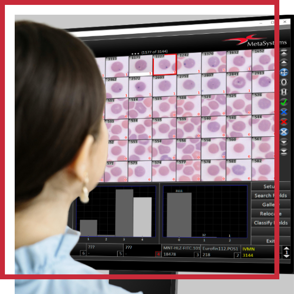
The in vivo MN test (rodent erythrocyte MN assay) normally uses bone marrow or peripheral blood of rodents which have been treated with the test substance. The red blood cells from the sample are classified as polychromatic (PCE; immature) or normochromatic (NCE; mature) erythrocytes. In PCE, the main nuclei have been extruded, and MN remain behind in the otherwise anucleated cytoplasm. An increase in the frequency of micronucleated polychromatic erythrocytes in treated animals is an indication of induced chromosome damage. PCE and NCE are distinguished by color (after applying a May-Gruenwald-Giemsa stain), and the ratio between both populations bears information about cell cycle delays due to toxic side effects of the testing agent. It is important to know that scoring automation of the in vivo MN test requires samples prepared from column purified cell solutions, and that it is recommended to prepare samples with cyto-centrifugation. Only this way of preparing slides assures reliable and error-free automated analysis.
Automation of the in vivo MN test with the Metafer software is done in a completely unattended workflow. The scanning system operated by Metafer SCAN automatically acquires images of the sample and analyzes the intensity range of the staining. With reference to the staining quality, PCE and NCE are automatically distinguished based on their color. The scanning workflow is robust enough to cope with varying staining qualities and with different ratios of PCE and NCE (for instance, if peripheral blood is used instead of bone marrow). Once PCE and NCE colors are defined, each cell is assigned to one of the classes, and micronuclei are detected with Metafer MetaCyte. Typically, more than 1,000 cells per minute are scanned and processed.
Automation of the in vivo MN test with Metafer MetaCyte is fully compliant with the respective OECD guideline (OECD guideline #474), and with the GLP regulation framework.


