Spot Counting
RapidScore: Streamlining Automated Image Acquisition and Expert Knowledge
- Unattended and automated imaging of interphase nuclei and FISH signals with focus stacking.
- Enhanced expert review with RapidScore empowers experts to efficiently evaluate data.
- Swift confirmation or modification of spot pattern suggestions using the RapidScore keyboard.
- Supports blinded review for multiple evaluators among team members.
- Adaptable to various probes, preparation methods, and color channels.
- Outcome presentation in an image gallery alongside comprehensive data tables and graphs.
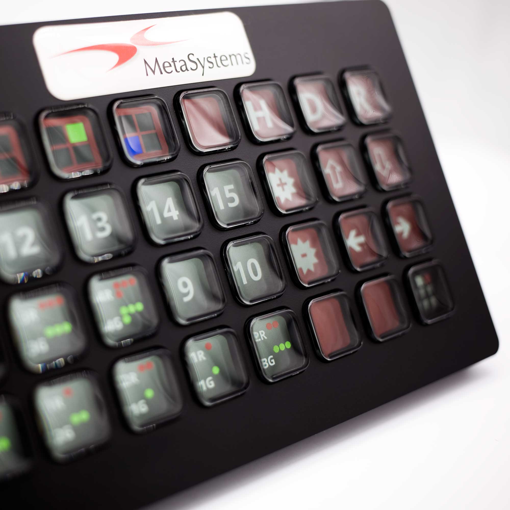
The Metafer Customization Package Spot Counting leverages the power of automation to automate tedious tasks, reducing manual labor and ensuring consistency in the evaluation of FISH signals. By incorporating advanced technology and tailored configurations, laboratories can optimize their processes, save time and improve overall productivity. The system automatically captures cell nuclei and FISH signals, enabling focus stacking for imaging signals on various focal planes. The outcome comprises a compilation of nucleus images presented in a Gallery format, along with spot pattern recommendations, and a summary display through graphical representations and tables.
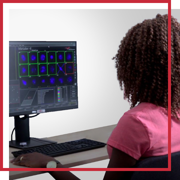
A cornerstone of the Metafer Customization Package Spot Counting is the RapidScore review process, which empowers experts to efficiently evaluate data. The RapidScore keyboard showcases spot patterns specific to the probe layout on the keys. Pressing a key allows for the confirmation or modification of Metafer suggestions, and the associated nucleus is marked as reviewed. If the user identifies any extra signal patterns not shown on the keyboard during the assessment, these can be effortlessly included in the evaluation process.
When involving multiple evaluators per slide, results blinding becomes crucial. Metafer addresses this with the CellReview window, displaying a processed and an unprocessed image of the nucleus together with representations of the individual color channels. An overview display provides the context around the nucleus. After a set number of evaluated nuclei, the file can be transferred to the next evaluator (up to five independent evaluators), ensuring blinded results to prevent influence.
The traditional manual evaluation method is time-consuming, repetitive, prone to errors, and often conducted in low-light conditions, leading to fatigue and decreased accuracy. Additionally, there's a lack of comprehensive image documentation and checks according to the four-eyes principle. MetaSystems proposes a workflow that integrates automation and human expertise to streamline FISH spot counting. Configurations created as part of this package aim to reduce processing time and enhance lab efficiency. The Customization Package Spot Counting provides a flexible workflow that is adaptable to various locus-specific probes. It seamlessly manages diverse probe layouts, preparation methods, and color channels, allowing for a smooth transition to digital signal counting.
Search Information Files (SIFs) are generated either by your compatible LIMS or manually in the Metafer software. SIFs may contain data on slide quantity, FISH probe specifics (including layouts like Dual Color Break Apart, Dual Fusion, etc.), and hybridization patterns on the individual specimens. Each slide can be recognized by a barcode and is linked to its respective case and culture. Metafer identifies the slides, either by their bar codes or by manual input, loads the according SIF, and executes the scans. It automatically captures cell nuclei and FISH signals, generating a compilation of nucleus images, spot pattern recommendations, and a summary display.
RapidScore is a crucial step in the workflow that allows experts to efficiently evaluate data by focusing on areas of interest and identifying anomalies. This process combines automation and human insight to draw well-informed conclusions from the data. The RapidScore keyboard displays anticipated spot patterns specific to the probe layout on its keys, allowing experts to swiftly confirm or modify spot suggestions with a keystroke. Experts can easily include unlisted patterns during evaluation.
The Image Gallery displays identified nuclei alongside FISH signals, allowing users to zoom in and navigate through focal planes. Data is categorized and presented in tables and graphs, with a summary of expert-validated outcomes for each nucleus.
Once data collection is complete and the cytogenetics experts have concluded their review, multiple avenues exist for presenting the outcomes. Neon incorporates an inherent Reporting Interface tailored precisely for this function, streamlining the generation of individualized and visually captivating reports. Furthermore, data can be effortlessly extracted from Neon — such as returning it to the LIMS — or combined into comprehensive statistical queries to facilitate summarization.
MetaSystems software provides, among other functions, features to assist users with image processing. These include, but are not limited to, the use of machine and deep learning algorithms for pattern recognition. The output generated in this process should be regarded as preliminary suggestions and, in any case, mandatorily requires review and assessment by trained experts.
MetaSystems offers Customization Packages for application workflows that have been successfully implemented for customer labs using standard Metafer platform functionality. It is expected that they can be implemented for other customer labs using similar workflows and slide preparation procedures. If a Customization Package is purchased, MetaSystems product specialists will – based on their experience from other similar application cases - support the customer lab in adapting the Metafer software configuration to their needs. The performance of the solution will depend on the quality of the customer slides and the expertise of the users, MetaSystems cannot specify or guarantee any performance parameters. The validation of the solution for clinical use is the sole responsibility of the customer lab.


