Clinical Cytogenetics, Cancer Genetics and Cell Biology
- Cytogenetics
- Hematology
- Oncology
- Immunology
- Rare Cell Detection
With the responsibility for patients on one hand, growing demand and increasing costs on the other, physicians responsible for cytogenetics and cancer genetics institutes find themselves pulled between these two extremes. But with automation of the tedious, yet critical image analysis, MetaSystems provides the tools to take the pressure off and helps to maintain, or even increase, quality standards with a much shorter case turnaround time.
In laboratory environments, data typically comes from many different, and often unrelated, sources. For example, patient data may be provided by a central laboratory information system (LIS), specimen samples at the lab bench may be identified by bar codes, images are generated by automated scanning systems or interactive capture stations, and external files may need to be kept.
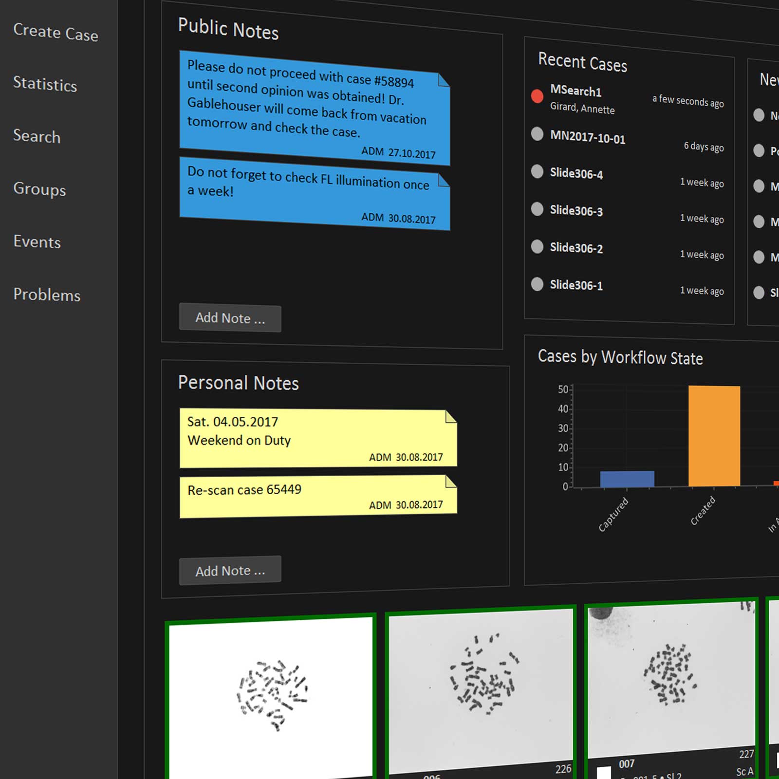
The case and image management software Neon, which is part of the latest Ikaros and Metafer software versions, has been designed to integrate and organize all of these data elements, and provide highly efficient tools to manage results. Neon revolutionizes laboratory workflow management by using an innovative, secure file format and organizing and displaying information in an extremely efficient and customizable manner. Users always have easy access to precise information about case status, history of images, analysis results, and more. Data import and export, even with customized data fields and global cross-application reporting, is available to effectively manage daily workflow.
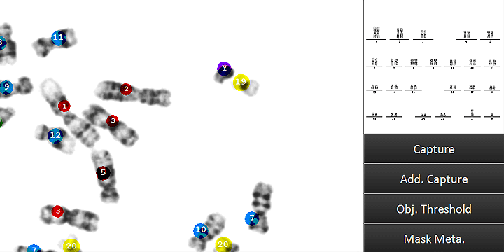
Automated imaging is a core competence of MetaSystems. Metafer, our globally renowned software, manages fully automated slide scanning. The Metafer based scanning system handles automated location of metaphases and acquisition of high-resolution images without user input. The images may be analyzed on any workstation running the Ikaros Karyo M module. Ikaros Karyo M can process metaphase images automatically captured with an integrated imaging system managed by Metafer. The optimized screen architecture of Ikaros Karyo M allows for extremely fast and efficient interactive on-screen karyotyping. Ikaros Karyo M works with any chromosome banding method, with fluorescence, and with chromosomes of any species. Smaller labs may configure a manual system equipped with Ikaros BASE M, a suitable microscope, PC, and camera to facilitate manual image acquisition.
Ikaros Karyo C is the perfect tool for interactive color fluorescence analysis. Ikaros Karyo C uses images that were automatically acquired with a Metafer based smart scanning solution. With Ikaros Karyo C, users can enhance, process, and display fluorescence in situ hybridization (FISH) results. In addition, Ikaros Karyo C also supports multicolor FISH (mFISH) and multicolor chromosome banding (mBAND). A manual imaging system operated with Ikaros BASE C can also manage the acquisition of up to 12 color channels.
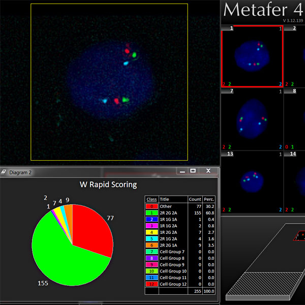
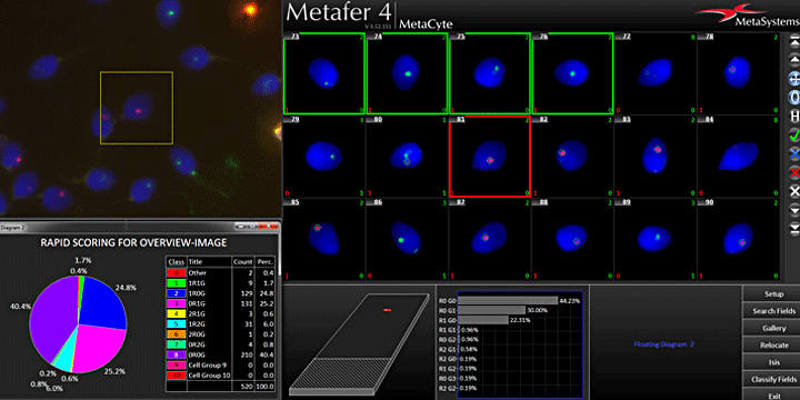
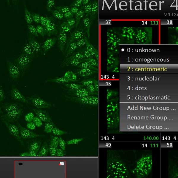
Our sophisticated image analysis algorithms can automatically process FISH slides for spot counting through the Metafer MetaCyte module. Metafer is well-adapted to the continuously growing portfolio of DNA probes from our partner company, MetaSystems Probes. Smart Scanning algorithms (e.g., an optimization of the scan based on cell density) guarantee a fast and robust workflow. The possibility to set up advanced scanning protocols allows an external LIMS to operate the Metafer based smart slide scanning remotely. Once the automated scanning is completed, all detected cells are displayed in an easily-navigable gallery, and detected signal patterns are summarized in convenient graphs and tables in Metafer MetaCyte.
The highly efficient RapidScore workflow allows for combining manual scoring strategies with the advantages of automated analysis. With RapidScore, cells that have been automatically pre-classified can easily be revised and, if required, re-classified with just one keystroke. With the external dynamic keypad for RapidScore, cell classes and the most important navigation features are directly accessible. New classes of cells can be dynamically defined based on signal and fusion patterns. Comprehensive graphs are updated on-the-fly during the scoring procedure. RapidScore also supports blind studies with multiple scorers.
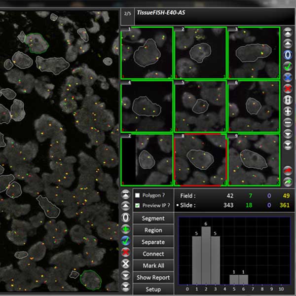
Innovative imaging solutions by MetaSystems are also suited to apply automated FISH imaging workflows to tissue sections. In this sense, they complement DNA FISH probes of MetaSystems Probes, which are optimized for tissues. The many tools under the hood of the Metafer based microscope imaging allow for creating extremely sophisticated Smart Scanning procedures for the analysis of FISH signals in selected regions of tissue:
- LIMS sends a Search Information File (SIF) to the Metafer operated scanning system to start the smart scanning of tissue samples.
- VSlide generates a slide overview image from a tissue sample, for example stained with H&E stain. The resulting images are sent online to the pathologist.
- The pathologist manually selects regions of interest in the overview image.
- Selection data is sent back to the Metafer operated scanning system, which matches the regions of interest to the DAPI image of a FISH labeled sample from the same specimen.
- All color channels and multiple focus planes of the regions are automatically captured using high power objectives.
- Resulting images are sent to the scorers. Cells are automatically pre-segmented.
- In Metafer's dedicated TissueFISH program surface, scorers pick the cells for analysis with the mouse; cells are immediately analyzed with Metafer's scoring algorithms for expert review.
- Results are automatically exported to LIMS or summarized in user-adaptable reports.
Autoimmune diseases are caused by abnormal immune responses against the constituents of the organism. As a result, inflammations or even severe organ damages can arise. Autoantibodies are located in the blood serum of the patient. Their presence is significant for the study of autoimmune diseases in preclinical phase, therefore they are employed in screening programs aimed at identifying individuals at risk.
The smart scanning solution based on the Metafer software enables routine analysis of fluorescence autoimmune samples in a highly reproducible way. Guaranteed constant lighting conditions thanks to LED illumination, well-defined integration parameters, flexibility in the use of high-throughput multi-field specimen, and powerful image processing algorithms are the key features of the Metafer based smart scanning solution to automatically score autoimmune samples. The unrivaled scanning speed allows digitizing whole specimen in just a few minutes. Bidirectional communication with third-party middleware or LIMS, and the sophisticated Search Information File (SIF) driven scanning workflow guarantees seamless integration with existing procedures. Interactive on-screen analysis of cell patterns is carried out with the help of automated image processing algorithms in Metafer MetaCyte. Gallery images of results, precise relocation of objects on the slide, and comprehensive data summary guarantee full control over the results.
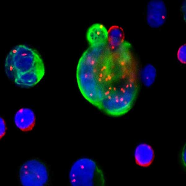
Circulating tumor cells (CTCs) are cells that have shed into the vasculature or lymphatics from a primary tumor and are carried around the body in the circulation. CTCs thus can be considered as seeds for the subsequent growth of additional tumors (metastases). The clinical relevance of CTC detection lies in the prediction of prognosis and survival, in monitoring therapy efficacy and potential relapse, and also in supervising drug resistances in real time.
There are several known methods for isolation and/or enrichment of CTC from peripheral blood. Even in enriched samples, however, manual detection and analysis of CTCs can take up to several hours, which renders the method inappropriate as a routine test. The smart scanning based on the Metafer software with its capability to combine different analysis strategies in a single scan has the power to overcome these limitations. A typical analysis of a CTC sample takes much less than an hour, and can be done considering several different markers applied to the same cell:
- DAPI signal to obtain morphology data, e.g., to pre-select CTC candidates using their sizes and/or shapes as criteria.
- Epithelial markers such as EpCAM, CK8, CK18, CK19.
- Mesenchymal markers such as Vimentin and Twist.
- Centromeric FISH signals to identify aneuploidies.
The manifold options to gate, sort and revise the population of detected cells facilitate quick check and full traceability of results.
Metafer 4.3 and Ikaros 6.3 are classified as in vitro diagnostic medical devices (IVD) in the European Union in accordance with In Vitro Diagnostics Regulation (EU) 2017/746 or In Vitro Diagnostic Medical Device Directive 98/79/EC, respectively, and carry the CE label unless otherwise indicated. Use all MetaSystems IVD products only within the scope of their intended purpose.
MetaSystems products are used in many countries worldwide. Depending on the regulations of the respective country or region, some products may not be used for clinical diagnostics.
Some hardware components supplied by other manufacturers are not included in MetaSystems IVD products and are therefore not IVD medical devices.
Please contact us for further information.