Pathology and Tissue Imaging
- Sample Digitization
- Digital Pathology and FISH
- Telepathology
MetaSystems has designed a smart scanning solution for FISH imaging and visualization tools to be integrated in the routine workflow of pathology labs. Smart slide scanning, networking, and documentation of results help to generate faster and better diagnoses.

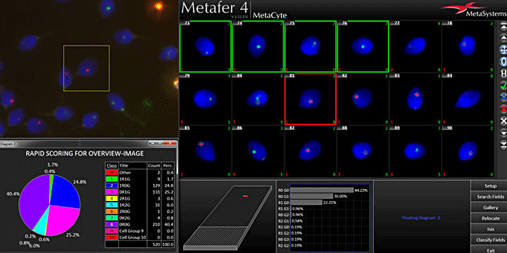
The renowned Metafer based scanning solution combines the advantages of a motorized microscope with modern, high-quality imaging automation. Therefore, the Metafer based scanning system is not restricted to certain magnifications or contrasting modes. Even Z-stacks can automatically be acquired, thus, providing the possibility to virtually 'focus' through the digitized sample.
Sample digitization with Metafer and VSlide is very easy. The Metafer operated scanning system automatically reads and interprets bar code labels and uses the encoded information to perform smart, multi-stage scanning workflows. Alternatively, dedicated imaging protocols can be provided from external software, e.g., by a LIMS (laboratory information management system). All images are accessible immediately after scanning and can be opened on any workstation with the free viewer software.
Though the virtual slides are stitched from individual image fields to achieve an unrivaled image quality, the scanning is extremely fast. A combination of a high-resolution camera, ultra-fast scanning hardware, and the high-capacity SlideFeeder x80 make the Metafer operated scanning a fast, yet precise scanning.
The flexibility of the Metafer software predestines the operated scanning system for being used for complex and particular imaging tasks, within pathology and beyond. No matter whether diatom images from the sea floor shall be visualized and identified, or fruit fly larvae are to be detected and digitized using a combination of Nomarski phase contrast and fluorescence – there is almost no conceivable imaging application the Metafer operated smart scanning could not cover.

Based on the wishes of users and lab managers, MetaSystems has designed a sophisticated scanning solution to analyze FISH signals in tissue sections. Core of the MetaSystems solution is a combination of dedicated DNA probes (MetaSystems Probes‘ XL range of locus-specific FISH probes target amplifications, deletions, or translocations of gene regions involved in solid tumors) and an innovative scanning system operated by the Metafer software.
The analysis workflow is based on the powerful analysis engine of Metafer, extended with a new, convenient analysis environment. The user interface supports either conventional mouse control or the optional interactive pen display.
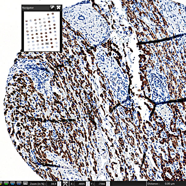
As initial step a digital brightfield, e.g., H&E slide is captured. The pathologist calls up the digital slide and selects on screen the tumor region on the virtual slide that needs to be FISH-scored.
Next, the FISH slide (from a subsequent section of the same block) is being scanned at low magnification to generate an overview. Displayed side by side to the marked digital H&E image, the tumor region can easily be transferred to the FISH slide.
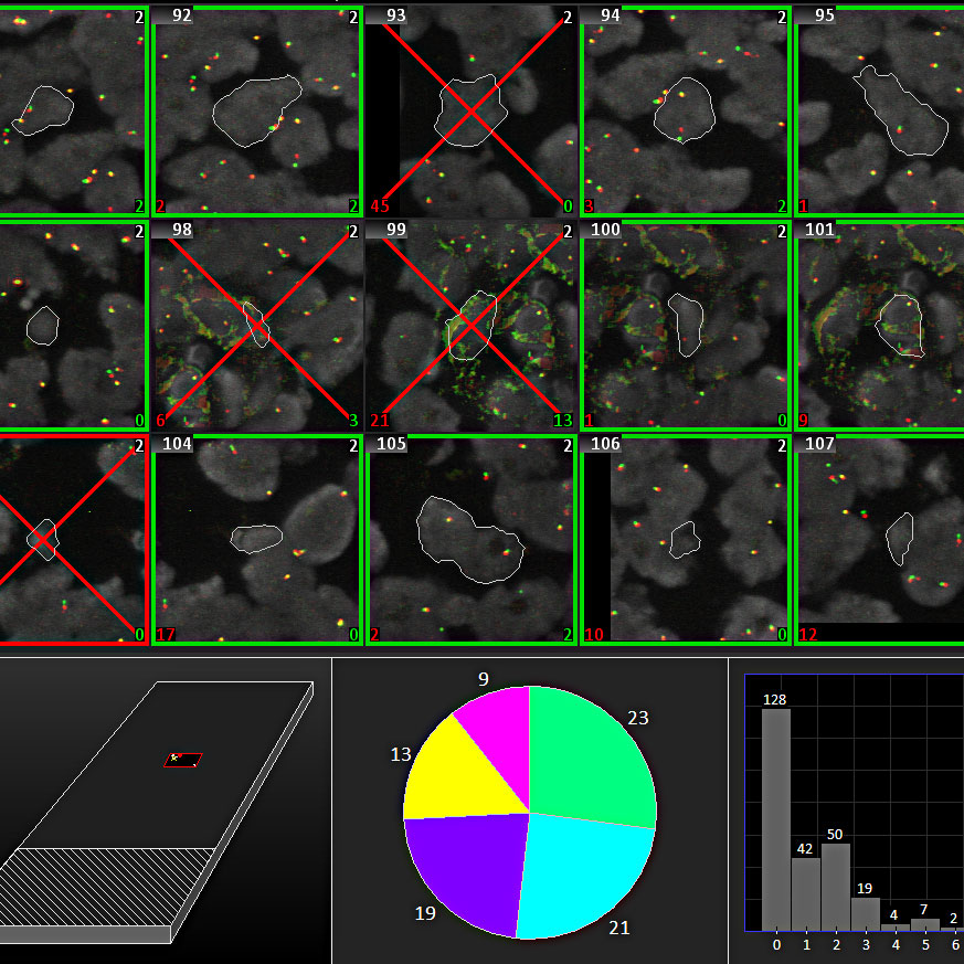
The Metafer operated scanning system now has all the information to start automatic image acquisition of the FISH slide at higher magnification. Cell nuclei that are isolated or slightly connected will be separated automatically and spot-counted. Manual tools for segmentation help to separate touching nuclei for immediate automatic scoring until the preset number of cells to be analyzed has been reached. The Metafer TissueFISH module can be easily set up to match the individual analysis standards. For instance, it is possible to define a minimum number of cells to be analyzed, and also to define a number of independent readers.
For final review, the Metafer TissueFISH presents a full synopsis to the pathologist, comprising the cell gallery with the scoring results, the virtual DAPI slide showing the positions of analyzed cells, and the corresponding H&E virtual slide. Every cell can be traced back to the tissue section to confirm its location within the preselected tumor region. Final results can either be exported as raw data, allowing for being processed in external software, or be summarized in comprehensive, user-adaptable reports.

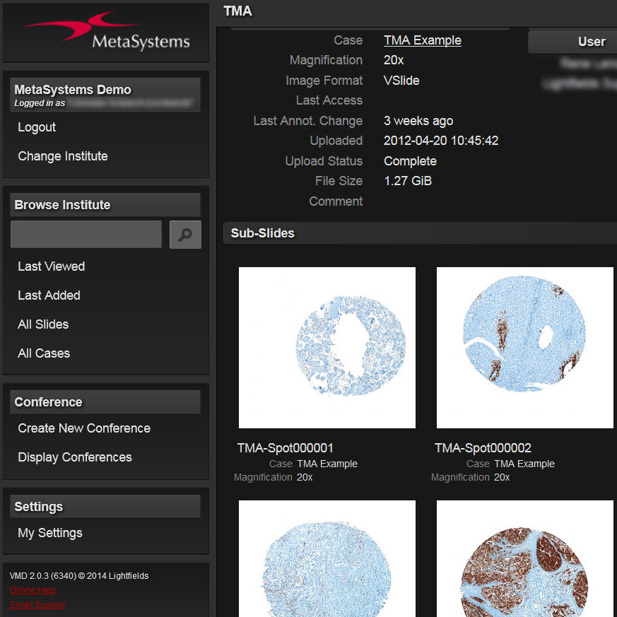
Once a pathology sample is digitized, it can be easily shared with others. The Metafer image file format makes this even more convenient: it contains a lot of additional information, e.g. on focus planes, on analyzed cells and signals, on annotations, and much more. Thus, any colleague who has access to the file, and who is opening the image in the viewer, can see all this information. Additionally, important image and meta data are stored digitally, and can be retrieved anytime for inspection.
Metafer 4.3 and Ikaros 6.3 are classified as in vitro diagnostic medical devices (IVD) in the European Union in accordance with In Vitro Diagnostics Regulation (EU) 2017/746 or In Vitro Diagnostic Medical Device Directive 98/79/EC, respectively, and carry the CE label unless otherwise indicated. Use all MetaSystems IVD products only within the scope of their intended purpose.
MetaSystems products are used in many countries worldwide. Depending on the regulations of the respective country or region, some products may not be used for clinical diagnostics.
Some hardware components supplied by other manufacturers are not included in MetaSystems IVD products and are therefore not IVD medical devices.
Please contact us for further information.


