More Applications
Your application does not match any of the other categories? MetaSystems may anyway be able to find a solution for your imaging problem.
Smart imaging solutions by MetaSystems excel through a unique combination of flexibility and reliability. This makes it easy to adapt a scanning system operated by the Metafer software to new applications. Please see below some success stories - maybe your application can also be automated?
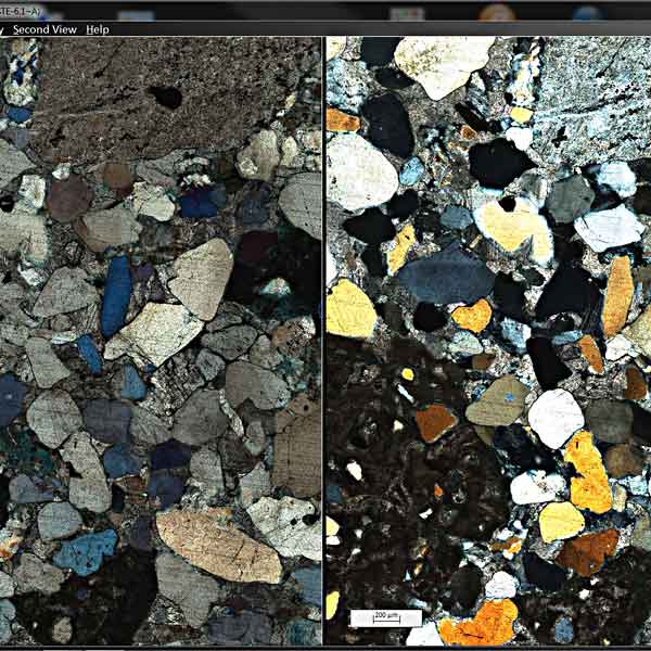
Digital microscope based imaging has changed the way the findings of petrographic examinations are nowadays carried out, documented and presented. Moreover, automation of such studies offers huge improvements in productivity and potentially significant reductions in the unit costs of examinations. Automated high-throughput digitization of geo-materials using several microscopic techniques that are routine in this field currently represents a substantial step forward in this sense.
MetaSystems has now set up a smart scanning solution for unattended scanning of geological samples. The Metafer operated scanning system offers a unique combination of outstanding scanning hardware with high-performance imaging software. Therefore, it constitutes an extremely versatile and robust workflow for microscopy automation.
MetaSystems has set up the first scanning solution for the digitization of geological samples that can be used for a high variety of contrasting methods and illumination techniques. This makes the solution designed by MetaSystems a genuine, all-in-one imaging solution for petrography, no matter whether translucent thin rock sections shall be observed with plane- and/or crossed-polarized transmitted light, whether block-mounted, opaque and polished metal specimens shall be inspected using reflected light illumination, or whether coal samples are assessed with epi-fluorescence illumination.
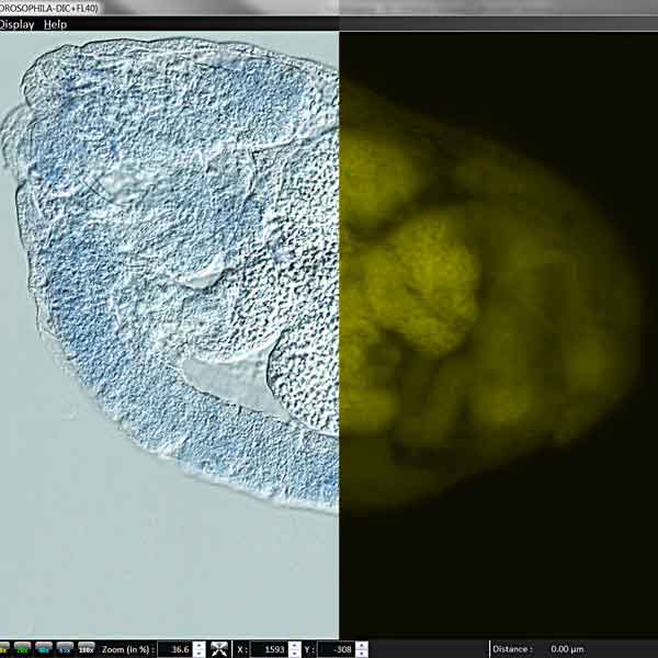
To visualize RNA expression in Drosophila larvae, the central imaging facility of a large biotechnology lab wanted to combine Nomarski phase contrast images with fluorescence. Since the Metafer software has the flexibility to support any contrasting method the microscope offers, it was easily possible to create an adapted workflow. The resulting images contain a color image of the Drosophila larvae acquired with Nomarski phase contrast, and a fluorescence channel showing the spatial distribution of RNA expression in the larvae.
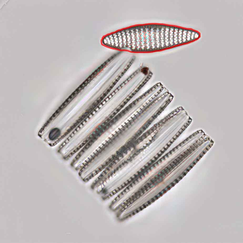
A few years ago, MetaSystems was approached by a renowned marine research institute. The request involved imaging, and identifying diatoms in samples obtained from the sea floor and other places. Diatoms, a major group of algae, are important indicators for environmental conditions. Diatoms are enclosed with a cell wall made of silica showing a large diversity in appearance. A Metafer operated scanning software and an adapted viewer software were installed and configured to communicate with existing diatom identification software. Now, the high-quality diatom images from Metafer are used routinely as a basis for automated diatom detection and identification.
Arabidopsis thaliana is a 20-25 cm tall flowering plant native to Europe, Asia, and northwestern Africa with a rapid life cycle of six weeks. It is used extensively as a model organism in plant biology and genetics. With about 157 million base pairs and five chromosomes, Arabidopsis has one of the smallest genomes among plants, and it was the first one to be sequenced in 2000.
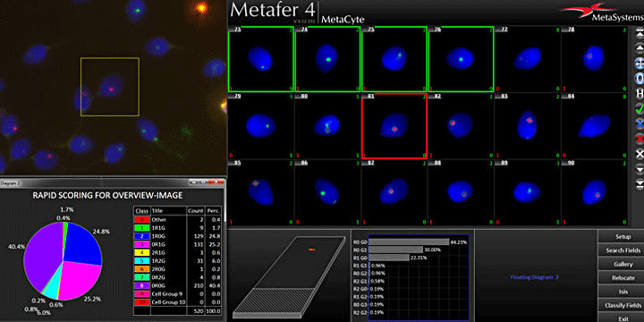
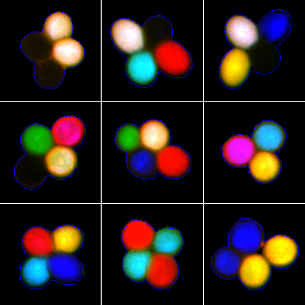
It is known that two A. thaliana genes, QRT1 and QRT2, are required for pollen separation during normal development. In certain mutants, however, pollen grains are released in tetrads. A tetrad is a cluster of 4 pollen grains that are the 4 products of one meiosis and which are still attached together. In a visual assay utilizing transgenic marker constructs it is possible to encode pollen-expressed fluorescent proteins of three colors in the quartet mutant background. This bears the possibility to study the results of one single meiosis and to analyze the crossing over interference (inhibition of nearby crossing overs) based on the specific fluorescence color pattern of the tetrads.
After harvesting, however, the fluorescence of the fluorescent reporter proteins in the tetrads vanishes within 4 hours. Therefore, the number of analyzed tetrads is limited if manual microscopy is used. An automated imaging protocol based on the smart scanning operated by the Metafer software, however, is capable of finding and identifying tetrads on the sample automatically. Thus, far more tetrads can be analyzed in the time before the markers vanish. Analysis of the tetrads is also automated by classifying the different color patterns. All data are conveniently summarized in automated reports.
Metafer 4.3 and Ikaros 6.3 are classified as in vitro diagnostic medical devices (IVD) in the European Union in accordance with In Vitro Diagnostics Regulation (EU) 2017/746 or In Vitro Diagnostic Medical Device Directive 98/79/EC, respectively, and carry the CE label unless otherwise indicated. Use all MetaSystems IVD products only within the scope of their intended purpose.
MetaSystems products are used in many countries worldwide. Depending on the regulations of the respective country or region, some products may not be used for clinical diagnostics.
Some hardware components supplied by other manufacturers are not included in MetaSystems IVD products and are therefore not IVD medical devices.
Please contact us for further information.