Forensics
- Forensic Sperm Detection
- Forensic Histopathology
Forensic scientists are tasked with the collection, preservation, and analysis of scientific evidence during the course of a criminal investigation. Within the field of forensic sciences, detection of semen is the primary method to confirm sexual assault. Other evidence, such as tissue samples, are used to examine the nature of incidents that may be linked to a particular crime.
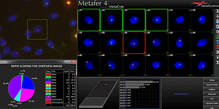
Sperm Cell Finding in Forensic Specimen
Driven by AI
- Replacing lengthy and tedious manual interaction with automated slide scanning.
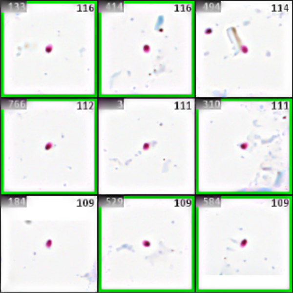
- Processing of standard forensic samples stained with “Christmas Tree” (Nuclear Fast Red/Picroindigocarmine) or Baecchi stain (acid fuchsin and methylene blue).
- Circa 15 minutes scanning time per slide with suitable specimen and hardware.
- Unattended slide scanning overnight possible (circa 40 – 50 slides o/n).
- Easy review of sperm cells with convenient image gallery of the Metafer software.
- Storage of all coordinates of detected sperm cells and quick relocation of objects.
- Easy transfer of object coordinates to laser micro-dissection systems.
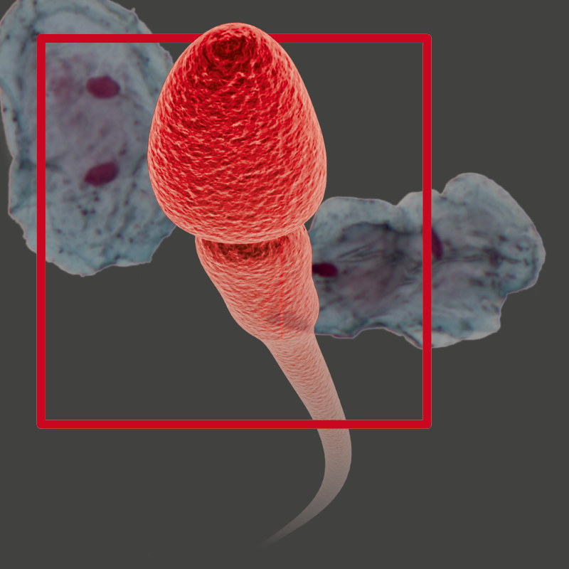
Sperm cells are a crucial piece of evidence for solving sexual crimes and are usually detected by microscopy. The smart scanning solution by MetaSystems automates the search for sperm cells in forensic specimens. Automation of microscopy imaging can significantly contribute to reduce turnaround times in specimen examination.
In some cases of sexual offenses, the detection of semen is a crucial piece of evidence. The success of this method depends on many pre-analytical and analytical procedures. In the laboratory, identification of archived sperm cells is done by visual inspection, after which sufficient quantities of sperm cells must be separated from other cells to obtain male DNA profiles. The manual procedure is tedious, time-consuming, and error-prone. As a result, many forensic laboratories criticize a backlog, and the time to solve a crime might be greatly delayed by this alone.
MetaSystems offers a reliable smart scanning solution to automatically locate sperm cells. An advanced algorithm based on a Deep Neural Network (DNN) allows for a highly sensitive detection of sperm cells with the Metafer SpermFinder DNN module. Detected sperm cells can easily be inspected on screen with the convenient Metafer software display. In addition, the positions of sperm cells on the slides are saved and can be transferred to a compatible laser micro-dissection system for targeted extraction of the detected sperm cells.
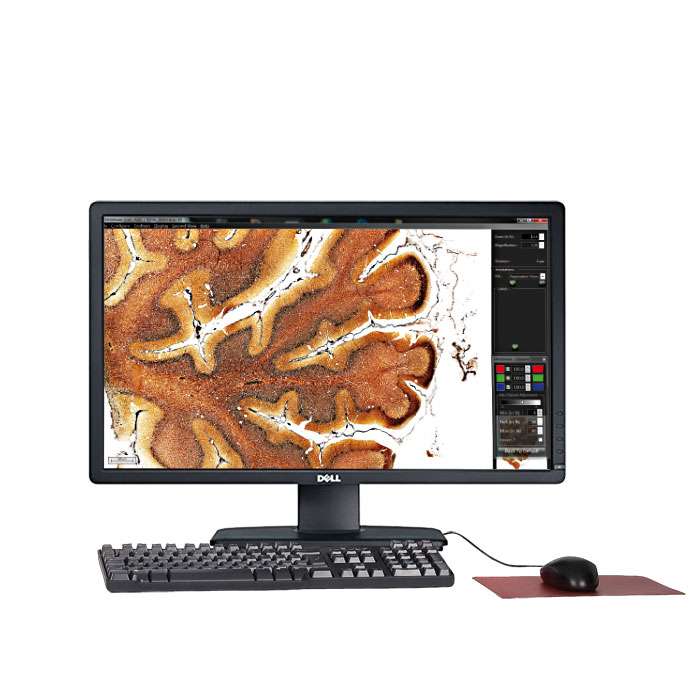
Forensic histology or histopathology applies histological techniques to forensic pathology practice. It becomes very useful in the case of a sudden death where trauma and poisoning need to be ruled out. Microscopic examination of human tissue can yield valuable information about the nature and extent of an injury present in an individual that may be related to the cause of death. Findings can be quite significant because the examiner will pass this information on to those responsible for the criminal investigation and may also need to testify in court. This is why quick responses, the 4-eyes principle for any assessment, and a thorough documentation of the findings become very important here.
The Metafer operated smart scanning and VSlide system provide an extremely versatile imaging toolbox offering many features helping to achieve these scopes. Samples of various origins can be fully digitized and subsequently used as basis for interpretation. Once digitized, degradation of the sample is of no importance anymore – the evidence is at hand whenever it is needed. Furthermore, it now becomes very easy to discuss over the image, to share opinions, and to develop findings.
The outstanding flexibility of VSlide provides a completely new insight into the sample. Regular brightfield light microscopy can easily be combined with fluorescence, with phase contrast, and even polarized light microscopy. Images obtained with these different methods can be superimposed, and cross-evidence can be visualized. Once elaborated, any investigation protocol can be made highly reproducible. This helps to reduce subjectivity, and to move closer towards robust QC/QA conditions.
Metafer 4.3 and Ikaros 6.3 are classified as in vitro diagnostic medical devices (IVD) in the European Union in accordance with In Vitro Diagnostics Regulation (EU) 2017/746 or In Vitro Diagnostic Medical Device Directive 98/79/EC, respectively, and carry the CE label unless otherwise indicated. Use all MetaSystems IVD products only within the scope of their intended purpose.
MetaSystems products are used in many countries worldwide. Depending on the regulations of the respective country or region, some products may not be used for clinical diagnostics.
Some hardware components supplied by other manufacturers are not included in MetaSystems IVD products and are therefore not IVD medical devices.
Please contact us for further information.